
Human eye can see more shades of Green than any other colour

Did you know that pirates used to wear earrings because they thought that they improved their eyesight? Brown coloured eyes are the most in the world. The surgical elimination of the brown pigment can help people have blue eyes. Naturally occurring blue coloured eyes are quite rare. Interestingly the human eye, a photosensitive sense organ, is the most complex organ of the human body after the brain. The human eye can see millions of shades of different colours and identify them however it can see most shades of green. This is simply because of human eye evolution, there were forests all around and hence predators could attack from all directions in the forest. Hence the human eye needed to have a better site of different shades of green. Come, let’s dig deeper into the structure and function of the Human Eye.
Table of Contents
Structure and Function of the Human Eye
The eyebrows do not serve any particular biological function but still cannot be categorised as a vestigial organ. This is because with the help of eyebrows we can communicate and express our feelings. Apart from this, they also perform the role of keeping away liquid (such as sweat) from the eyes.
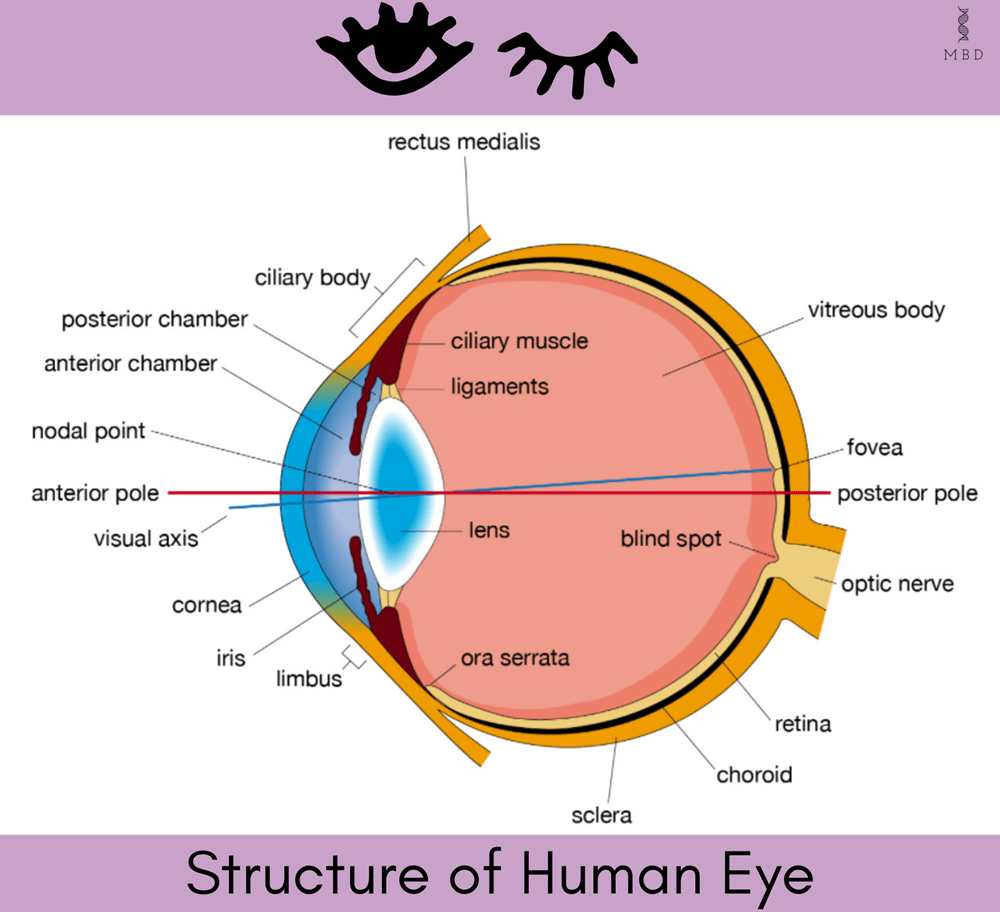
Image Source: britannica.com
The human eye helps us to see the world around us because of the optic nerve that connects the eyes to the brain. With the help of this optic nerve, the brain can interpret the signals from the photosensitive cells in the eyes. In order to understand the mechanism of vision, it is very crucial to understand the structure and function of the Human Eye.
In Pop Culture references, we always say that ‘the gateway to the soul of every individual is through their eyes’.
Around the eye, a nictitating membrane is present, called Plica semilunaris. It is a vestigial organ (the organ that does not serve any particular function) that has the role to protect the eyes when underwater. Since humans are terrestrial animals we do not require the nictitating membrane. Each eye is present in its orbit. The human eye has two eyelids: the upper eyelid and the lower eyelid. The eyelids bear eyelashes too. The white coloured portion of the eye that we can see is the sclera. The coloured portion of the eye is due to pigmentation in the iris and the black dot that we can observe is the pupil.
Eye Layers
The human eye, a photosensitive sensory organ, is a spherical structure. The eyeball has 3 layers.
Outer Layer
The outermost layer, which is the external layer, mainly consists of fibrous tissue. The outermost layer can be divided into two: the sclera and the cornea. The 5/6 portion of the outermost layer is the sclera, the white portion of our eye. 1/6 portion of the eye is the cornea. The cornea is an avascular outermost layer of the eye that consists of simple stratified squamous epithelium tissue.
Since the cornea does not have any blood vessels, it can easily be transplanted without even blood group matching or any other issue. The sclera is mainly composed of dense fibrous connective tissues. The cornea is the anterior portion whereas the rest of the posterior is the sclera.
Middle Layer
The middle layer of the eye has many blood vessels, unlike the outer layer which is avascular. Because of the presence of blood vessels, the middle layer looks bluish. The middle layer mainly consists of a choroid. The 2/3 posterior area of the middle layer is a very thin choroid. The anterior portion as seen in the diagram becomes thick and forms the ciliary body. The ciliary body consists of ciliary muscles and can be termed as a thick choroid. The main function of the ciliary body is accommodation. The ciliary body further continues to form the iris. Iris is ectodermal in origin and it has pigments. Iris is an opaque structure and can be termed as the visible coloured portion of the human eye.
Depending upon the pigment present in Iris the colour of the eye is determined. The iris holds the crystalline lens of the eye. This crystalline transparent lens is held in place by ligaments that are present on the ciliary body. The pupil is the aperture between the iris. The iris consists of fibre that contains dilators and constructive muscles. The iris helps in regulating the size of the pupil. The pupil aperture determines how much light will enter the eye.
Inner Layer
The innermost layer of the eye is called the retina. The retina is the place where image formation will take place after light will enter through the eye. The retina consists of three layers of cells. They are ganglion cells, bipolar cells and photoreceptor cells. The photoreceptor cells are present just after the retina. The photoreceptor cells will further on send the signals to bipolar cells and ganglion cells to relay it to the central nervous system.
The blind spot is a region in the retina where no photoreceptor cells are present hence no image is formed at the blind spot. The position where the optic nerve leaves the eye and blood vessels enter, the blind spot is present. Macular luteya, a yellowish pigmented spot, is present at the posterior pole of the human eye just lateral to the blind spot. It has a central pit called Fovea. Fovea has only cones present. Due to the dense packing of cones at this region, the resolution of the image that is generated by a retina is the highest. The retina has two types of photoreceptor cells called rods and cones. Rods help us see in dim light whereas cones help in daytime vision.
Aqueous chamber and Vitreous chamber
The aqueous chamber and vitreous chamber are two important components of the human eye. The space between the cornea and the lens that can be seen in the diagram is filled with Aqueous humour, a thin watery fluid that can be replaced. This chamber is called an Aqueous chamber. The space between the lens and retina is called the Vitreous chamber. The vitreous chamber is filled with the transparent gel called vitreous humour which is non-replaceable. Vitreous humour is responsible for maintaining the shaping, pressure and refractive index of the eye. Aqueous humour and Vitreous humour are transferred to the human eye with the help of the canal of schlemm.

Image Source: aao.org
Mechanism of Vision
After understanding the structure and function of the Human Eye, let us now understand how the human eye helps us to see the colourful world around us. To understand the mechanism of vision, we will have to understand more about the photoreceptor cells. The retina has two types of photoreceptor cells termed rods and cones. 130 million rods are present whereas 7 million cones are present. Rods are the photosensitive structures that help us in dim vision whereas Cones help us see colour. Photopic vision is the daylight vision and the colour vision as well, this is the function of cones. The scotopic vision that is the Twilight vision is the function of rods.
Rods consist of rhodopsin. Rhodopsin can also be termed as visual purple. It is a purplish red coloured protein that is a derivative of vitamin A. Vitamin A derivatives are formed from carotene, aldehyde. In the human eye, characteristic photopigments are present that respond to the primary colours red, green and blue. If the human eye wants to process different types of colours they are formed by the mixture of these. When all the primary colours are stimulated equally white light sensation is produced.
What happens?
The mechanism of vision starts when visible electromagnetic waves first pass through the cornea. then the pupil regulates the amount of light that will enter the eye. If the light is too much then the pupil constricts and if we are in dim light the pupil dilates. This is the reason when we come out of cinema halls we have difficulty in our vision for some time. As the light enters through the cornea the light goes to the crystalline transparent lens. This lens generates an image on the retina. The retina has photoreceptor cells called rods and cones. These photopigments consist of a photosensitive compound called rhodopsin. Rhodopsin is composed of opsin and retinal. Opsin is a protein and retinal is an aldehyde of vitamin A.

Image Source: researchgate.net
Whenever an image is formed on the retina, the retinal is dissociated from opsin. As this dissociation occurs an action potential is generated because of the change in structure. Due to action potential generation membrane permeability changes. This signal is first received by bipolar cells (as the name indicates the cells have an axon and one dendrite). The bipolar cells transmit these nerve impulses to the ganglion cells. These action potentials are then transmitted to the optic nerve which takes it directly to the visual cortex area of the brain, the central nervous system. Depending upon existing information and memory in the brain, the visual cortex recognises it and interprets the image and gives us this sensation of vision.
Disorders of the Human Eye
If we look closely at the human eye, a photosensitive sensory organ and slight blood vessel-like structures are present on the outside. This is the conjunctiva. Inflammation can occur in conjunctiva which is termed conjunctivitis. In some situations, vitreous humour and aqueous humour channel called canal of schlemm can be blocked and Kala motia can occur. It can also cause glaucoma i.e. permanent blindness. There is no cure for Kala motia only further damage can be prevented. Sometimes due to vitamin A deficiency night blindness can occur because if Vitamin A is deficient then retinal cannot be produced and therefore the rods cannot function properly and scotopic vision gets interfered with.
The crystalline transparent lens is responsible for the accommodation. the human eye can see between 25 cm to infinity. The lens helps adjust its focal length to help us see all through this range. As we grow old the power of ciliary muscles for accommodation decreases due to genetic reasons.

Image Source: teachoo.com
Myopia and hypermetropia are common disorders that are observed. Myopia is short-sightedness whereas hypermetropia is far-sightedness. In the case of myopia because the crystalline transparent lens is not getting adjusted properly the image is being formed just before the retina and in the case of hypermetropia, the images are formed behind the retina. Myopia hypermetropia can be corrected with the help of lenses or spectacles. Do make sure to consult your doctors if you have any problems in your vision.
Hope the article helped you get some idea about the structure and function of the Human Eye.
Keep reading at MBD to find out more about other interesting sensory organs.
Team MBD
Also check out- Awareness of STDs and AIDS – My Biology Dictionary

