
Types of staining techniques in Microbiology
In today’s time research industry is booming as never before, all thanks to SARS-CoV-2. Do you understand the need to study microbes? To begin with, microorganisms are versatile and play a major role in various biochemical processes such as epidemiology, biotechnology, climate change, food spoilage, etc. In order to study bacteria, archaea, viruses, algae, and prions, we need to stain them before we study and observe them under a microscope since they differ greatly in their structure and characteristics. The main purpose behind staining is to visualize morphological characteristics and increase visibility and preserve microorganisms. Staining is the most important technique used by scientists in the field of microbiology. Stains are all about beautiful colours, let’s have a look at the types of staining techniques used by microbiologists as an effective tool.
Table of Contents
Stain & Staining
A stain is an organic compound that contains a benzene ring with an auxochrome and a chromophore group.
- Auxochrome: A chemical compound that conveys the property of ionization of chromogen and binds to fibre or tissues.
- Chromophore: A chemical group that helps to dye our subject by imparting colour to benzene.
Staining is simply a technique to visualize cells better under a microscope so that one can observe and study about cell’s morphology and chemical nature of the cell under observation.
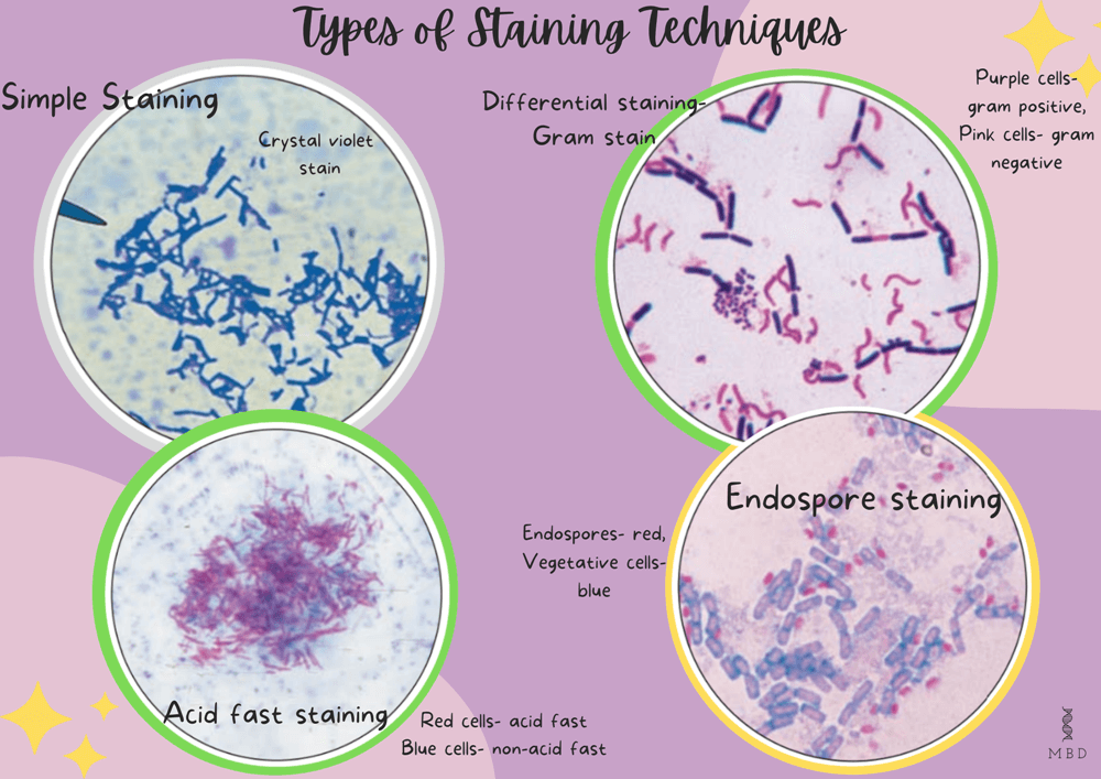
Types of staining Techniques in Microbiology
- Simple staining: Staining using a single stain.
- Differential staining: Staining using two or more contrasting stains.
- Staining of specific cell structures.
Image Source: Prescott’s Microbiology 7th Edition
Simple staining
- To begin with, a method of staining that uses only one single dye/stain.
- There is only one staining step and the whole subject is stained with the same color.
- Another key concept is that we cannot differentiate organisms using simple staining.
- Simple staining is particularly used to stain the whole cell or specific cellular components.
- Microorganisms often can be stained very satisfactorily by simple staining as it is very easy to use and has a simple process.
Types of a simple staining
There are further two types of simple staining-
Direct/ positive staining
It stains objects (in a single colour) not the background. These are basic dyes and have positively charged groups.
Examples– Crystal violet, Basic Fuchsin.
Indirect/negative staining
It stains only the background. These are acidic dyes, because of their negative charged groups.
Example- Acid fuchsin, Rose Bengal.
Differential staining
- Secondly, in the differential staining process, we use more than one chemical stain.
- By using more than one chemical stain one can better differentiate between different microorganisms or cellular components in a single subject/organism.
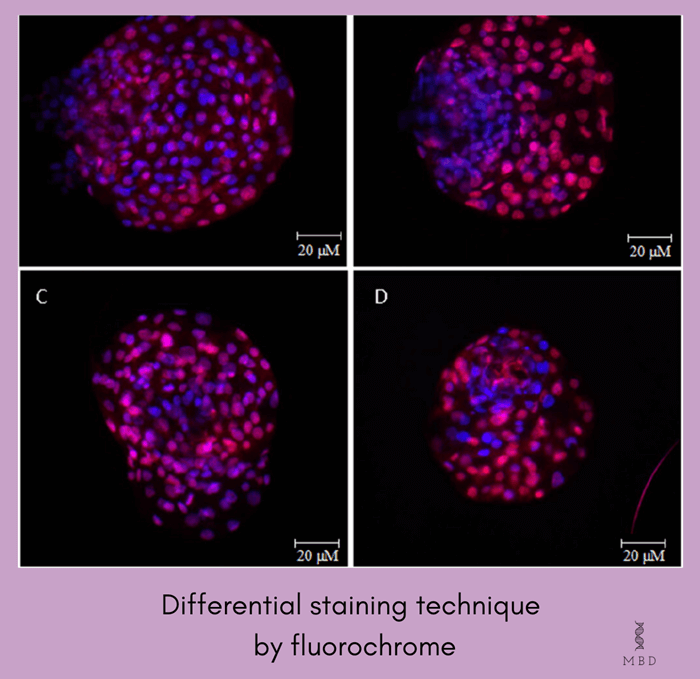
Image Source: Garcia et al (www.researchgate.net)
Types of differential staining
Gram staining
- Gram stain was developed by Christian gram (Danish physician) in 1884, it is the most widely employed staining method in bacteriology
- This staining procedure particularly defines two bacterial groups, Gram-positive (positive by gram method), and Gram-negative (negative by gram method). This is the starting point for the bacterial identification procedure.
- Moreover, Gram-positive bacteria have thick, dense, relatively, non-porous walls, while gram-negative have thin walls with lipid-rich membranes.
Requirements for Gram staining
Gram staining is a four-part procedure. The specimen is heat-fixed and mounted on the slide, in the first part. The reagents required:
a) Crystal violet- acts as a primary stain
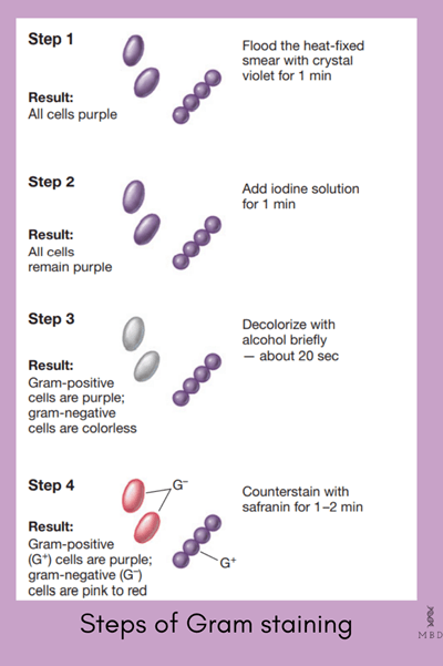
Image Source: Brock Biology of microorganisms
b) Iodine solution acts as the mordent
c) Ethanol- as a decolourizer
d) Safranin- the counter stain
Procedure of Gram staining:
- In the first step, both gram-positive and negative bacteria are fixed on a slide and stained with the primary stain(basic dye crystal violet), both appear purple after completion of this step.
- Then bacteria are particularly treated with mordant (iodine solution), which helps the stain to retain better by making a violet-iodine complex known as “iodine lake”. The end colour is purple after this step.
- This is the differential step, Gram’s decolourizer ( Ethanol with acetone) is added to the specimen, at the end of this step Gram-positive bacteria retain their purple colour while gram-negative decolorizes.
- In the final step, a basic dye ( safranin) is applied. Since the gram+ bacteria retain the purple colour they remain unaffected, while the gram- bacteria get stained with the basic dye and appear pink.
- At the end of this step, finally, the gram-positive appears purple and the gram-negative appears pink.
Acid-fast staining
- Another important differential staining most commonly used to identify Mycobacterium tuberculosis and M. Leprae.
- From ‘ACID FAST’, we can understand that once stained the acid-fast bacteria resist the decolourization with an acidified organic solvent.
- Mycobacteria has very lipid-rich cell walls majorly made up of mycolic acid (a specialized group of branched-chain hydroxy lipids).
- Additionally, it is also called the Ziehl-Neelsen method.

Image Source: Taylor, G. M., Stewart, G. R., Cooke, M., Chaplin, S., Ladva, S., Kirkup, J., … & Young, D. B. (2003). Koch’s Bacillus–a look at the first isolate of Mycobacterium tuberculosis from a modern perspective. Microbiology, 149(11), 3213-3220.
Procedure of Acid-fast staining:
- To perform acid-fast staining, the specimen is heat-fixed and the primary stain (carbol fuchsin) is added, while continuous is provided to slide over a steam bath.
- The heat helps melt the waxy barrier and allows the absorption of the dye by the cell.
- Then the slide is allowed to cool down and then a decolourizer (solution of alcohol and acid) is added.
- The acid-fast cells particularly resist the decolourization and retain the primary stain and the other remaining cells get decolourized, then methylene blue is used as a counterstain.
- In the end, finally, the acid-fast bacteria appears a bright pink colour and all other cells appear blue.
You can’t miss checking out this one– Mechanism of Immune Response – My Biology Dictionary
Staining of specific cell structures
Many special staining procedures have been developed to study specific structures under a light microscope.
Capsule staining
- A technique that reveals the presence of capsules, a network usually made of polysaccharides that surrounds many bacteria and some fungi.
- Due to the non-ionic nature of the capsule, it doesn’t get stained by an acidic stain, but a basic stain like Crystal violet stains the cell as well as the capsule.

Image Source: Loh, J., Lorenz, N., Tsai, C. J. Y., Khemlani, A. H. J., & Proft, T. (2017). Mucosal vaccination with pili from Group A Streptococcus expressed on Lactococcus lactis generates protective immune responses. Scientific reports, 7(1), 1-9.
Procedure of capsule staining:
- Being hypertonic, Copper sulfate solution causes diffusion of the stain towards the outer surface of the cell.
- After drying the slide, the stain which is not passed from the capsular layer during diffusion remains in the capsular layer.
- Copper sulfate then decolourizes the capsule.
- The capsule particularly appears as a faint blue halo around a purple cell.
Endospore staining
- Like acid-fast staining, this also requires harsh treatment to dye the target structure significantly.
- An endospore is an exceptionally resistant structure produced by some bacterial genera
- Endospores are particularly meant to survive under unfavourable conditions for a long period, so they are not easy to stain.
- Endospores are not stained well by most dyes, but once the spore is stained, they strongly resist decolourization. This property is the basis of most endospore staining methods

Image Source: Brock Biology of Microorganisms
- A specialized process called Schaeffer-Fulton procedure is used for staining endospores.

Procedure of endospore staining:
Following are the steps of endospore staining-
- The bacteria is heated and stained using the primary stain( malachite green), being a very strong stain it gets to penetrate the endospores.
- In the next step, the rest of the cell is washed with water (free of dye) and is counter-stained using safranin (secondary stain).
- Finally, this yields a green endospore and while the vegetative cells appear red in color of safranin.
To conclude, we are too fascinated by the types of staining and their beautiful colourations. How about you?
Stay tuned! Thank you for reading.
Team MBD
Watch how to perform Gram staining- Gram Staining Procedure Animation Microbiology – Principle, Procedure, Interpretation – YouTube

