Phagocytosis – Principle, Mechanism and Process

Phagocytosis refers to the process in which a cell engulfs a large particle by extending its plasma membrane. This gives rise to an internal compartment called the phagosome inside which the particle is trapped. Phagocytosis consists of the recognition and ingestion of particles larger than 0.5 μm. The cells that perform phagocytosis are called phagocytes. Phagocytes can ingest microbial pathogens and thus contribute to the clearance of billions of cells that are turned over every day in an organism. Therefore phagocytosis becomes biologically significant not only for microbial elimination but also for tissue homeostasis. In this article, we will throw some light on the principle and mechanism of phagocytosis.
Also check out- Enthralling structure of tRNA (transfer RNA) – My Biology Dictionary

Image source: www.creative-biolabs.com
Did you know that cells can eat up?
Table of Contents
Principle Of Phagocytosis
The principle of phagocytosis is based on the destruction of microorganisms. Phagocytosis is a non-specific defense mechanism in which various phagocytes engulf and destroy the disease-causing microorganisms. Phagocytes also initiate the processes of the immune system for eliminating the invaded antigen. The basic principle one can say about phagocytosis is that the microorganisms are recognized by surface receptors on the phagocytes that engulf the microbe and a phagosome is formed. In this way, the disease-causing microbe or waste cell debris is destroyed in the body.
The Mechanism Of Phagocytosis
Despite the strong defenses of our protective epithelial layers, some pathogens have evolved strategies to penetrate these defenses, and epithelia may be disrupted by wounds, abrasions, and insect bites that may transmit pathogens. Phagocytic cells make up the next line of defense against pathogens penetrating the epithelial cell barriers. Phagocytosis is one of the main mechanisms of the innate immune defense mechanism. It is one of the first processes of responding to an infection. Although most cells are capable of phagocytosis, some cell types perform it as an immune response to kill the pathogen. These are called ‘professional phagocytes.’ Thus phagocytosis is old in evolutionary terms, even in invertebrates and microorganisms.
What Initiates Phagocytosis?
Did you think about how a phagocytic cell recognizes microbes, triggering their phagocytosis?
There are receptors present on the surface of phagocytes that directly recognize specific conserved molecular components on the surfaces of microbes. These components could be protein parts or any fragment of the microorganism. These conserved molecular components are on the surfaces of microbes, such as cell wall components of bacteria, fungi, etc. Therefore these components usually present in many copies on the surface of a bacterium, fungal cells, parasite, or virus particle, are called pathogen-associated molecular patterns (PAMPs).
The phagocytes improve interactions of receptors with possible targets,
(i) by creating active membrane protrusions that allow the cell to explore larger areas, increasing the chances for receptors to engage their ligands, and
(ii) by selectively removing some of these larger glycoproteins allowing the receptors to diffuse more freely on the membrane.
Process Of Phagocytosis
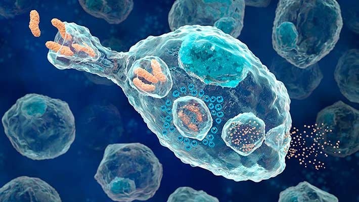
The process of phagocytosis begins with the attachment and ingestion of microbial particles into a bubble-like structure called a phagosome.
- After recognition of the pathogen, the membranous extensions called pseudopodia arise and move towards the pathogen as a result of chemotaxis.
- The pathogen becomes attached to these membrane evaginations and it keeps on extending until the pathogen gets completely engulfed by the membrane.
- The ingested pathogen is in the phagosome that fuses with the lysosome. This, therefore, results in a phagolysosome.
- Once inside the phagocyte, the phagosome containing the microorganism fused lysosome. It contributes to enzymes that destroyed the bacterium or the pathogen.
- These lysosomal enzymes kill the pathogen and after that, the digestion products are released from the cell or rejected as waste.
Flow cytometry: History, Components, Applications- MBD (mybiologydictionary.com)
The process of phagocytosis is very complex and we have only a partial understanding of it. However, several important steps directed by actin remodeling, to form the pseudopodia to cover the particle, can be identified. First, the membrane-associated cytoskeleton, of the resting phagocyte, needs to be disrupted so that it can extend. Second, nucleation of actin filaments takes place in order to initiate F-actin polymerization and extension of membrane for pseudopodia.
The Phagosome
The organelle formed by phagocytosis of a material is called the phagosome. After its formation, it moves towards the center of the phagocytes and fuses with lysosome forming phagolysosome to get hydrolytic enzymes for the degradation of the trapped pathogen. Progressively, the phagolysosome is a package of acidified, activating degradative enzymes.
The degradation in the phagosome is in two ways: Oxygen-dependent and Oxygen independent.
The Phagosome Formation
Phagocytic receptors with ligands on the surface of target particles commence phagosome formation. Then, receptors must aggregate to initiate signaling pathways that regulate the actin cytoskeleton in the cell, so that the phagocyte can produce membrane protrusions for engulfing the particle. At last, the particle is enclosed in a new vesicle that pinches out from the plasma membrane.
How Are The Pgahocytosed Microbes Killed In Phagocytosis
- The binding of microbes like bacteria, fungi, protozoan parasites, and viruses to phagocytes via pattern recognition receptors or opsonins and opsonin receptors activates signaling pathways.
- These signals trigger Actin polymerization, resulting in membrane extensions around the microbe particles. The phagosomes then fuse with lysosomes forming phagolysosomes and, in neutrophils, with preformed primary and secondary granules.
- It contains an arsenal of antimicrobial agents that then kill and degrade the internalized microbes. Here these agents include acid-activated enzymes, and hydrolytic enzymes, which contain an arsenal of antimicrobial agents that then kill and degrade the internalized microbes.
Also check out- The Centrifuge – Definition, Principle, Types and Applications (mybiologydictionary.com)
An important additional activity of macrophages in the spleen and in the liver especially Kupffer cells is to recognize, phagocytose, and degrade ageing and damaged red blood cells. As these cells age, novel molecules that are recognized by phagocytes accumulate in their plasma membrane. Phosphatidyl serine flips from the inner leaflet to the outer leaflet of the lipid bilayer and is recognized by phosphatidyl serine receptors on phagocytes. Modifications of erythrocyte membrane proteins have also been detected that may promote phagocytosis.
Phagocytosis is thus an important phenomenon for the killing and destruction of such microbes. Also, the waste cell debris gets destroyed there and there’s no such accumulation of it in the body. Therefore phagocytosis is a significant phenomenon and biological process for the body. It is a natural predictor for such invading microorganisms in the body. The Principle Of Phagocytosis is a bit complex to understand. Hope this article was useful to you.


Nice.
Awesome!!